Anamorphic video

The term anamorphic refers to a distorted image that appears normal when viewed with an appropriate lens. When shooting film or video, an anamorphic lens can be used to squeeze a wide image onto a standard 4:3 aspect ratio frame. During projection or playback, the image must be unsqueezed, stretching the image back to its original aspect ratio.

By default, 16:9 anamorphic video displayed on an standard monitor appears horizontally squeezed, meaning images look tall and thin. The advantage of this was in the past that producers could shoot wide-screen material using inexpensive equipment. Rescaling anamorphic video in order to see the entire wide screen frame on a standard definition 4:3 monitor is called letterboxing, and results in the loss of the maximum resolution available in the source footage. A wide screen (16:9) allows video-makers more room for creativity in their shot composition.

To check the support of anamorphic videos by different players, I created three mp4 videos from scratch, based on squeezed test pictures :

testbild_anamorphic

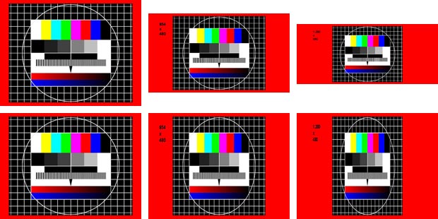
Source pictures  640×480, 854×480 and 1.280×480 squeezed to 640×480 pictures

The following ffmpeg script creates a video from a squeezed source image towards a stretched widescreen video with a ratio 2.35:1.

ffmpeg ^
-loop 1 ^
-f image2 ^
-i testbild_2_35_1_squeezed.jpg ^
-r pal ^
-vcodec libx264 ^
-aspect 235:100 ^
-crf 23 ^
-preset medium ^
-profile:v baseline ^
-level 3.1 ^
-refs 1 ^
-t 30 ^
testbild_anamorphic_2_35_1.mp4
pause

The -aspect parameter handles the correct display aspect ratio (DAR). The MediaInfo tool shows that the video has 640×480 pixels, but an DAR of 2.35:1.
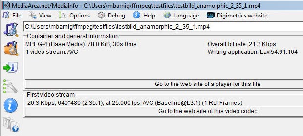
MediaInfo

MediaInfo

The VLC video player stretches the video based on the DAR. Videos with a wrong DAR in the metadata can be resized manually by changing the aspect ratio in the corresponding video menu.
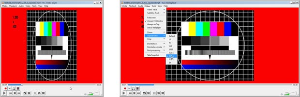
anamorphic video

VLC media player

More informations about anamorphic videos are available at the following links :

Human Brain Parts and Regions

Last update : October 11, 2014

human brain regions

brain regions

The brain is the center of the nervous system in all vertebrate and most invertebrate animals. From a philosophical point of view, what makes the brain special in comparison to other organs is that it forms the physical structure that generates the mind. Through much of history, the mind was thought to be separate from the brain. Even for present-day neuroscience, the mechanisms by which human brain activity gives rise to consciousness and thought remain very challenging to understand: despite rapid scientific progress, much about how the human brain works remains a mystery. The operations of individual brain cells are now understood in considerable detail, but the way they cooperate in ensembles of millions has been very difficult to decipher.

The human brain has three main parts :

  1. The cerebrum, or telencephalon (Grosshirn, cerveau), that fills up most of the skull, is involved in cognition and also controls movement.
  2. The cerebellum, or little brain (Kleinhirn, cervelet), that sits at the back of the head, under the cerebrum, controls coordination and balance.
  3. The brainstem (Hirnstamm, tronc cérébral), that sits beneath the cerebrum in front of the cerebellum, connects the brain to the spinal cord and controls automatic functions such as breathing, digestion, heart rate and blood pressure.

The human brain is divided into right and left halves (hemispheres). The left half controls movement on the body’s right side. The right half controls the body’s left side. In most people, the language area is mainly on the left. Preserved brains have a grey color, hence the name grey matter.

The brain’s wrinkled surface is a specialized outer layer of the cerebrum, called the cerebral cortex (what we see when we look at the brain). Each bump on the surface of the human brain is known as a gyrus, while each groove is known as a sulcus.

In a typical human the cerebral cortex is estimated to contain 15–33 billion neurons, each connected by synapses to several thousand other neurons. These neurons communicate with one another by means of long protoplasmic fibers called axons, which carry trains of signal pulses called action potentials to distant parts of the brain or body targeting specific recipient cells..

Traditionally the cerebral cortex is divided into four sections, which are known as lobes :

english latin deutsch français
Frontal Lobe Lobus frontalis Stirnlappen lobe frontal
Parietal lobe Lobus parietalis Scheitellappen lobe pariétal
Temporal lobe Lobus temporalis Schläfenlappen lobe temporal
Occipital lobe Lobus occipitalis Hinterhauptlappen lobe occipital

The Terminologia Anatomica (TA), the international standard on human anatomic terminology, developed by the Federative Committee on Anatomical Terminology (FCAT) and the International Federation of Associations of Anatomists (IFAA), released in 1998, defines two additional lobes : The limbic lobe, associated to emotion and memory and the insular cortex, associated to pain and some other senses.

The frontal lobe is associated with reasoning, motor skills, higher level cognition, and expressive language. The parietal lobe is associated with processing tactile sensory information such as pressure, touch, and pain. The temporal lobe is the location of the primary auditory cortex, which is important for interpreting sounds and the language we hear. The hippocampus is also located in the temporal lobe, which is why this portion of the brain is heavily associated with the formation of memories. The occipital lobe is associated with interpreting visual stimuli and information. The primary visual cortex, which receives and interprets information from the retinas of the eyes, is located in the occipital lobe.

The cerebral cortex is also segmented in cortical areas which are functionally or anatomically defined. Some examples are listed below :

human brain areas

brain areas

The brainstem is comprised of the hindbrain (rhombencephalon) and midbrain. The hindbrain contains structures including medulla oblongata, the pons and the reticular formation.

The limbic system contains glands which help relay emotions. Many hormonal responses that the body generates are initiated in this area. The limbic system includes the amygdala, hippocampus, hypothalamus and thalamus.

Great progresses in the analysis which parts of the brain are involved in a particular mental process have been made in the last years with the functional magnetic resonance imaging (fMRI).

More informations about human brain parts and regions are available at the following links:

at Wikipedia :

other sources :

BioBlender visualization

BioBlender is a software package built on the open-source 3D modeling software Blender. BioBlender version 1.0 for Windows and Linux was released on July 12, 2013. The first beta version of BioBlender (v 0.1) was presented in September 2010.

BioBlender

BioBlender

BioBlender is the result of a collaboration, driven by the SciVis group at the Institute of Clinical Physiology (CNR) in Pisa, Italy, between scientists of different disciplines (biology, chemistry, physics, computer sciences) and artists, using Blender in a rigorous but at the same time creative way.

With BioBlender users can handle proteins in the 3D space, displaying their surface in a photorealistic way, and elaborate protein movements on the basis of known conformations. Scientists all over the world study proteins at atomic level and deposit information in the public repository Protein Data Bank, where each molecule is described as the list of its atoms and their 3D coordinates.

BioBlender can be used for:

  • import and visualize Protein Data Bank (PDB) files (The PDB file format is a textual file format describing the three dimensional structures of molecules held in the Protein Data Bank)
  • simulate molecular dynamics and optimize protein motion
  • visualize complex protein surface properties  (e.g. MLP and EP surface properties)

A BioBlender tutorial was published by Raluca Andrei, Mike Chen Pan and Monica Zoppè, in the BlenderArt magazine N.31 in December 2010.

Biological and artificial neurons

Biological neurons

A biological neuron (nerve cell) is an electrically excitable cell that processes and transmits information through electrical and chemical signals. A chemical signal occurs via a synapse, a specialized connection with other cells. Neurons connect to each other to form neural networks. Neurons are the core components of the nervous system, which includes the brain, spinal cord, and peripheral ganglia. There are different types of neurons: sensory neurons, motor neurons and interneurons.

A typical neuron possesses a soma (perkaryon or cyton = cell body with nucleus), dendrites and an axon. Neurons do not undergo cell division.

Neurons

Neuron (Wikipedia)

Dendrites are thin structures that arise from the cell body, branching multiple times and giving rise to a complex dendritic tree. An axon is a special cellular extension that arises from the cell body and travels for long distances (as far as 1 meter in humans). The cell body of a neuron gives rise to multiple dendrites, but never to more than one axon, although the axon may branch hundreds of times before it terminates. The axon terminal contains synapses, specialized structures where neurotransmitter chemicals are released to communicate with target neurons. At the majority of synapses, signals are sent from the axon of one neuron to a dendrite of another, however there are a lot of exceptions.

All neurons are electrically excitable, maintaining voltage gradients across their membranes by means of metabolically driven ion (sodium, potassium, chloride, calcium) pumps. Changes in the cross-membrane voltage can alter the function of voltage-dependent ion channels. Each time the electrical potential inside the soma reaches a certain threshold, an all-or-none electrochemical pulse called an action potential is fired, which travels rapidly along the cell’s axon, and activates synaptic connections with other cells when it arrives.

Artificial neurons

An artificial neuron is a mathematical function conceived as an abstraction of biological neurons. The artificial neuron receives one or more inputs (representing the dendrites) and sums them to produce an output (representing the axon). Usually the sums of each node are weighted, and the sum is passed through a non-linear function known as an activation function or transfer function.

The first artificial neuron was the Threshold Logic Unit (TLU) first proposed by Warren McCulloch and Walter Pitts in 1943. This model is still the standard of reference in the field of neural networks and called a McCulloch–Pitts neuron. However, artificial neurons of simple types, such as the McCulloch–Pitts model, are sometimes characterized as caricature models, in that they are intended to reflect one or more neurophysiological observations, but without regard to realism.

In the 1980s computer scientist Carver Mead, who is widely regarded as the father of neuromorphic computing, demonstrated that sub-threshold CMOS circuits behave in a similar way to the ion-channel proteins in cell membranes. Ion channels, which shuttle electrically charged sodium and potassium atoms into and out of cells, are responsible for creating action potentials. Using sub-threshold domains mimicks action potentials with little power consumption.

At the Neuromorphic Cognitive Systems Institute of Neuroinformatics of the University of Zurich and ETH Zurich, a research group leaded by Giacomo Indiveri is currently developing, using the sub-threshold-domain principle, neuromorphic chips that have hundreds of artificial neurons and thousands of synapses between those neurons.

Volunteer Computing

Volunteer computing is an arrangement in which people (volunteers) provide computing resources to projects which use the resources to do distributed computing and/or storage. Distributed computing is a field of computer science that studies distributed systems. A distributed system is a software system in which components, located on networked computers, communicate and coordinate their actions by passing messages.

Neural networks are very good candidates for simulation by distributed computing systems because of their inherent parallelism and beacuse its simulation is a very time consuming process, due to the complex iterative process involved.

The first volunteer computing project was the Great Internet Mersenne Prime Search, which was started in January 1996. The term volunteer computing was coined by Luis F. G. Sarmenta, the developer of Bayanihan.

The Berkeley Open Infrastructure for Network Computing (BOINC) is the most widely-used middleware system for volunteer computing. It offers client software for Windows, Mac OS X, Linux, and other Unix variants. The project was founded at the University of California, Berkeley Space Sciences Laboratory, funded by the National Science Foundation. Other systems are XtremWebXgrid and Grid MP.

Volunteer computing systems must deal with the following problems, related to correctness :

  • Volunteers are unaccountable and essentially anonymous
  • Some volunteer computers occasionally malfunction and return incorrect results
  • Some volunteers intentionally return incorrect results or claim excessive credit for results

A list of distributed computing projects is provided at Wikipedia. Links to a few selected BOINC volunteer computing projects are listed below :

OpenWorm Caenorhabditis elegans

Last update : August 9, 2013

OpenWorm aims to build the first comprehensive computational model of the Caenorhabditis elegans (often called C. elegans, even if this term is a species abbreviation), a free-living, transparent nematode (roundworm), about 1 mm in length, that lives in temperate soil environments. With only a thousand cells, it solves basic problems such as feeding, mate-finding and predator avoidance.

OpenWorm background

Research into the molecular and developmental biology of C. elegans was begun in 1974 by Nobel prize laureate Sydney Brenner and it has since been used extensively as a model organism for development biology. Sydney Brenner founded the Molecular Sciences Institute in Berkeley, California.

Caenorhabditis elegans (Wikipedia)

Caenorhabditis elegans (Wikipedia)

The basic anatomy of C. elegans includes a mouth, pharynx, intestine, gonad, and collagenous cuticle. C. elegans has two sexes: hermaphrodites and males (0.05%).

C. elegans is one of the simplest organisms with a nervous system. In the hermaphrodite, this comprises 302 neurons whose pattern of connectivity (connectome) has been completely mapped and shown to be a small-world network. C. elegans was also the first multicellular organism to have its genome completely sequenced. The genome consists of six chromosomes (named I, II, III, IV, V and X) and a mitochondrial genome. The sequence was first published in 1998 with regular updates, because DNA sequencing is not an error-free process. The latest version released in the WormBase () is WS238.

WormBase is an international consortium of biologists and computer scientists dedicated to providing the research community with accurate, current, accessible information concerning the genetics, genomics and biology of C. elegans and related nematodes. Founded in 2000, the WormBase Consortium is led by Paul Sternberg of CalTech, Paul Kersey of the EBI, Matt Berriman of the Wellcome Trust Sanger Institute, Lincoln Stein of the Ontario Institute for Cancer Research, and John Spieth of the Washington University Genome Sequencing Center. Richard Durbin served as a principal investigator until 2010.

Additional informations about C. elegans are available at the following links :

  • WormBook – a free online compendium of all aspects of C. elegans biology
  • WormAtlas – an online database for behavioral and structural anatomy of C. elegans
  • WormClassroom – an education portal for C. elegans
  • WormImagethousands of unpublished electron micrographs and associated data
  • WormWeb.org – an interactive cell lineage and neural network
  • Cell Exlorer – a 3D visualization tool for the structural anatomy of C. elegans
  • C. elegans movies

OpenWorm open source project

Despite being extremely well studied in biology, the C. elegans still eludes a deep, principled understanding of its biology. The OpenWorm project uses a bottom-up approach, aimed at observing the worm behaviour emerge from a simulation of data derived from scientific experiments carried out over the past decade. To do so, the data available in the scientific community is incorporated into OpenWorm software models.

An open-source simulation platform called Geppetto is used by the OpenWorm Project to run these different models together. An OpenWorm Browser enables ready access to a cell-by-cell 3D representation of the nematode C. elegans in a WebGL enabled browser. The 3d browser was created with the help of the Google Labs Body Browser team. The browser has also been ported to an iOS app to support the project. All the code produced in the OpenWorm project is Open Source and available on GitHub.

The OpenWorm project is realized by a highly motivated group of individuals who believe in Open Science. The OpenWorm website includes a Blog, a Wiki, a FAQ and Donate page, lists about milestones, projects, events, publications, getting started and getting involved resources and more.

The core team members of the OpenWorm project are :

[jwplayer player=”1″ mediaid=”13386″]

Neuromorphic computing

neuromorphic computing by Spike Gerrell

credit : Spike Gerrell for the Economist

Neuromorphic computing is a concept developed by Carver Mead, describing the use of very-large-scale integration (VLSI) systems containing electronic analog circuits to mimic neuro-biological architectures present in the nervous system. Carver Mead is a key pioneer of modern microelectronics.

Today the term neuromorphic is used to describe analog, digital, and mixed-mode analog/digital VLSI and software systems that implement models of neural systems. Neuromorphic computing is a new interdisciplinary discipline that takes inspiration from biology, physics, mathematics, computer science and engineering to design artificial neural systems and autonomous robots, whose physical architecture and design principles are based on those of biological nervous systems.

The goal is to make computers more like brains and to design computers that have  features that brains have and computers do not have up to now :

  • low power consumption (human brains use about 20 watts)
  • fault tolerance (brains lose neurons all time without impact)
  • lack of need to be programmed (brains learn and change)

An important property of a real brain is that each neuron has tens of thousands of synaptic connections with other neurons, which form a sort of small-world network. Many neuromorphic chips use what is called a cross-bar architecture, a dense grid of wires, each of which is connected to a neuron at the periphery of the grid, to create this small-world network. Other chips employs what is called synaptic time multiplexing.

The Economist published a few days ago a great article “Neuromorphic computing – The machine of a new soul” with illustrations from the London-based illustrator Spike Gerrell.

Some neuromorphic computing reletad projects are listed below :

Neuromorphic computing is dominated by European researchers rather than American ones. The following links provide additional informations about neuromorphic computing related institutions and topics :

Artificial General Intelligence

Last update : August 7, 2013

Artificial General Intelligence (AGI) is an emerging research field aiming at the building of thinking machines; that is, general-purpose systems with intelligence comparable to that of the human mind (and perhaps ultimately well beyond human general intelligence). While this was the original goal of Artificial Intelligence (AI), the mainstream of AI research has turned toward domain-dependent and problem-specific solutions; therefore it has become necessary to use a new name to indicate research that still pursues the Grand AI Dream. Similar labels for this kind of research include Strong AI, Human-level AI, etc. Other AI researchers prefer the term of Synthetic Intelligence.

The research on AGI is interdisciplinary, focused on whole systems and includes scientific and philosophical investigation and software engineering.

Artificial General Intelligence Research Institute

The term AGI was first used by Mark Avrum Gubrud in November 1997. Fifty years after the launch of the Artificial Intelligence Project in Dartmouth in 1956, Ben Goertzel, Phil Goetz, Pei Wang and Bruce Klein organized the first Artificial General Intelligence Research Institute (AGIRI) workshop in May 2006 to bridge the gap between narrow AI and general-purpose AI. The AGI Research Institute was founded in 2001 with the mission to foster the creation of powerful and ethically positive Artificial General Intelligence. The institute is sponsored by Novamente LLC.

The aspects of Artificial General Intelligence are explained by Pei Wang and Ben Goertzel  in the introduction of their book Advances in Artificial General Intelligence (IOS Press, 2007).

The first conference on Artificial General Intelligence (AGI-08) was organized by AGIRI in March 2008 in Memphis, Tennessee, USA, in association with the Association for the Advancement of Artificial Intelligence (AAAI).

Artificial General Intelligence Society

Ben Goertzel, Pei Wang, Joscha Bach and others founded in September 2011 the Artificial General Intelligence Society (AGI society), a nonprofit organization with the following goals:

  • promote the study of artificial general intelligence (AGI), and the design of AGI systems
  • facilitate co-operation and communication among those interested in the study and pursuit of AGI
  • hold conferences and meetings for the communication of knowledge concerning AGI
  • produce publications regarding AGI research and development
  • publicize and disseminate by other means knowledge and views concerning AGI

The organization of the annual Artificial General Intelligence conference series, which was started in 2008 by AGIRI, has been taken over by the AGI society. The next conference (AGI-2013) will be held in Beijing, China, July 31 – August 3, 2013.

Some additional informations about AGI are available at the following links :

More links are provided in the updated post about Artificial Intelligence.

A look inside mice brains

A team of researchers at the Stanford University, lead by Mark Schnitzer, an associate professor of biology and applied physics, planted tiny probes inside mice brains to detect what were essentially mouse memories. The study was published February 10, 2013, in the online edition of Nature Neuroscience.

inside mice brains

Read a mouse’s mind

The experiment involved the insertion of a needlelike microscope into the hippocampus of the mice brains. The microscope detected cellular activity and broadcast digital images through a cell phone camera sensor that fit like a hat over the heads of the critters as they were running around. Over the course of a month, the scientists were able to document patterns of activity in about 1000 neurons of the mice brains where they store long-term information. To get the results, an engineered gene was injected into the mice brains so that their proteins were sensitive to calcium ions. That caused the magnified cells to light up on the computer screen in flashes of green fluorescence when the neurons were activated.

Three students, who worked on the project, have formed a startup company called Inscopix, and they plan to sell the technology to neuroscience researchers.

[jwplayer player=”1″ mediaid=”13056″]

More informations are available at the news website of Stanford University.

Synthetic Biology

Synthetic biology is the design and construction of new biological parts, devices, and systems, and the re-design of existing, natural biological systems for useful purposes. It combines biology and engineering with a focus on Biotechnology.

Synthetic biologists focus on finding how life works (the origin of life) or how to use it to benefit society, including the approach of biology by inserting man-made DNA into a living cell and the approach of chemistry by working on gene synthesis as an extension of synthetic chemistry.

The website syntheticbiology.org, originally started by a group of students, faculty and staff from MIT and Harvard, now regroups all individuals, groups and labs from various institutions who are committed to engineering biology in an open and ethical manner. The site is hosted on OpenWetWare and can be edited by all members of the Synthetic Biology community.

An exciting synthetic biology project was recently funded succesfully on Kickstarter : Glowing Plants: Natural Lighting with no Electricity. A few days ago, without explanation, Kickstarter quietly altered its guidelines for project creators, introducing a new term that bans creators from giving away genetically-modified organisms (GMOs) as rewards to their online backers (see the post Kickstarter bans project creators from giving away genetically-modified organisms edited by Duncan Geere at The Verge website).

More informations about synthetic biology are available at the following links :