Biological and artificial neurons

Biological neurons

A biological neuron (nerve cell) is an electrically excitable cell that processes and transmits information through electrical and chemical signals. A chemical signal occurs via a synapse, a specialized connection with other cells. Neurons connect to each other to form neural networks. Neurons are the core components of the nervous system, which includes the brain, spinal cord, and peripheral ganglia. There are different types of neurons: sensory neurons, motor neurons and interneurons.

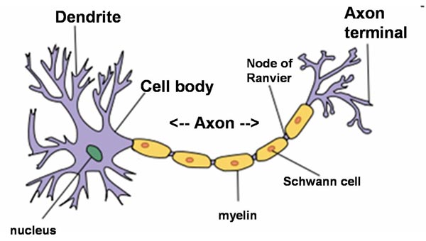
A typical neuron possesses a soma (perkaryon or cyton = cell body with nucleus), dendrites and an axon. Neurons do not undergo cell division.

Neurons

Neuron (Wikipedia)

Dendrites are thin structures that arise from the cell body, branching multiple times and giving rise to a complex dendritic tree. An axon is a special cellular extension that arises from the cell body and travels for long distances (as far as 1 meter in humans). The cell body of a neuron gives rise to multiple dendrites, but never to more than one axon, although the axon may branch hundreds of times before it terminates. The axon terminal contains synapses, specialized structures where neurotransmitter chemicals are released to communicate with target neurons. At the majority of synapses, signals are sent from the axon of one neuron to a dendrite of another, however there are a lot of exceptions.

All neurons are electrically excitable, maintaining voltage gradients across their membranes by means of metabolically driven ion (sodium, potassium, chloride, calcium) pumps. Changes in the cross-membrane voltage can alter the function of voltage-dependent ion channels. Each time the electrical potential inside the soma reaches a certain threshold, an all-or-none electrochemical pulse called an action potential is fired, which travels rapidly along the cell’s axon, and activates synaptic connections with other cells when it arrives.

Artificial neurons

An artificial neuron is a mathematical function conceived as an abstraction of biological neurons. The artificial neuron receives one or more inputs (representing the dendrites) and sums them to produce an output (representing the axon). Usually the sums of each node are weighted, and the sum is passed through a non-linear function known as an activation function or transfer function.

The first artificial neuron was the Threshold Logic Unit (TLU) first proposed by Warren McCulloch and Walter Pitts in 1943. This model is still the standard of reference in the field of neural networks and called a McCulloch–Pitts neuron. However, artificial neurons of simple types, such as the McCulloch–Pitts model, are sometimes characterized as caricature models, in that they are intended to reflect one or more neurophysiological observations, but without regard to realism.

In the 1980s computer scientist Carver Mead, who is widely regarded as the father of neuromorphic computing, demonstrated that sub-threshold CMOS circuits behave in a similar way to the ion-channel proteins in cell membranes. Ion channels, which shuttle electrically charged sodium and potassium atoms into and out of cells, are responsible for creating action potentials. Using sub-threshold domains mimicks action potentials with little power consumption.

At the Neuromorphic Cognitive Systems Institute of Neuroinformatics of the University of Zurich and ETH Zurich, a research group leaded by Giacomo Indiveri is currently developing, using the sub-threshold-domain principle, neuromorphic chips that have hundreds of artificial neurons and thousands of synapses between those neurons.

OpenWorm Caenorhabditis elegans

Last update : August 9, 2013

OpenWorm aims to build the first comprehensive computational model of the Caenorhabditis elegans (often called C. elegans, even if this term is a species abbreviation), a free-living, transparent nematode (roundworm), about 1 mm in length, that lives in temperate soil environments. With only a thousand cells, it solves basic problems such as feeding, mate-finding and predator avoidance.

OpenWorm background

Research into the molecular and developmental biology of C. elegans was begun in 1974 by Nobel prize laureate Sydney Brenner and it has since been used extensively as a model organism for development biology. Sydney Brenner founded the Molecular Sciences Institute in Berkeley, California.

Caenorhabditis elegans (Wikipedia)

Caenorhabditis elegans (Wikipedia)

The basic anatomy of C. elegans includes a mouth, pharynx, intestine, gonad, and collagenous cuticle. C. elegans has two sexes: hermaphrodites and males (0.05%).

C. elegans is one of the simplest organisms with a nervous system. In the hermaphrodite, this comprises 302 neurons whose pattern of connectivity (connectome) has been completely mapped and shown to be a small-world network. C. elegans was also the first multicellular organism to have its genome completely sequenced. The genome consists of six chromosomes (named I, II, III, IV, V and X) and a mitochondrial genome. The sequence was first published in 1998 with regular updates, because DNA sequencing is not an error-free process. The latest version released in the WormBase () is WS238.

WormBase is an international consortium of biologists and computer scientists dedicated to providing the research community with accurate, current, accessible information concerning the genetics, genomics and biology of C. elegans and related nematodes. Founded in 2000, the WormBase Consortium is led by Paul Sternberg of CalTech, Paul Kersey of the EBI, Matt Berriman of the Wellcome Trust Sanger Institute, Lincoln Stein of the Ontario Institute for Cancer Research, and John Spieth of the Washington University Genome Sequencing Center. Richard Durbin served as a principal investigator until 2010.

Additional informations about C. elegans are available at the following links :

  • WormBook – a free online compendium of all aspects of C. elegans biology
  • WormAtlas – an online database for behavioral and structural anatomy of C. elegans
  • WormClassroom – an education portal for C. elegans
  • WormImagethousands of unpublished electron micrographs and associated data
  • WormWeb.org – an interactive cell lineage and neural network
  • Cell Exlorer – a 3D visualization tool for the structural anatomy of C. elegans
  • C. elegans movies

OpenWorm open source project

Despite being extremely well studied in biology, the C. elegans still eludes a deep, principled understanding of its biology. The OpenWorm project uses a bottom-up approach, aimed at observing the worm behaviour emerge from a simulation of data derived from scientific experiments carried out over the past decade. To do so, the data available in the scientific community is incorporated into OpenWorm software models.

An open-source simulation platform called Geppetto is used by the OpenWorm Project to run these different models together. An OpenWorm Browser enables ready access to a cell-by-cell 3D representation of the nematode C. elegans in a WebGL enabled browser. The 3d browser was created with the help of the Google Labs Body Browser team. The browser has also been ported to an iOS app to support the project. All the code produced in the OpenWorm project is Open Source and available on GitHub.

The OpenWorm project is realized by a highly motivated group of individuals who believe in Open Science. The OpenWorm website includes a Blog, a Wiki, a FAQ and Donate page, lists about milestones, projects, events, publications, getting started and getting involved resources and more.

The core team members of the OpenWorm project are :

[jwplayer player=”1″ mediaid=”13386″]

Neuromorphic computing

neuromorphic computing by Spike Gerrell

credit : Spike Gerrell for the Economist

Neuromorphic computing is a concept developed by Carver Mead, describing the use of very-large-scale integration (VLSI) systems containing electronic analog circuits to mimic neuro-biological architectures present in the nervous system. Carver Mead is a key pioneer of modern microelectronics.

Today the term neuromorphic is used to describe analog, digital, and mixed-mode analog/digital VLSI and software systems that implement models of neural systems. Neuromorphic computing is a new interdisciplinary discipline that takes inspiration from biology, physics, mathematics, computer science and engineering to design artificial neural systems and autonomous robots, whose physical architecture and design principles are based on those of biological nervous systems.

The goal is to make computers more like brains and to design computers that have  features that brains have and computers do not have up to now :

  • low power consumption (human brains use about 20 watts)
  • fault tolerance (brains lose neurons all time without impact)
  • lack of need to be programmed (brains learn and change)

An important property of a real brain is that each neuron has tens of thousands of synaptic connections with other neurons, which form a sort of small-world network. Many neuromorphic chips use what is called a cross-bar architecture, a dense grid of wires, each of which is connected to a neuron at the periphery of the grid, to create this small-world network. Other chips employs what is called synaptic time multiplexing.

The Economist published a few days ago a great article “Neuromorphic computing – The machine of a new soul” with illustrations from the London-based illustrator Spike Gerrell.

Some neuromorphic computing reletad projects are listed below :

Neuromorphic computing is dominated by European researchers rather than American ones. The following links provide additional informations about neuromorphic computing related institutions and topics :

A look inside mice brains

A team of researchers at the Stanford University, lead by Mark Schnitzer, an associate professor of biology and applied physics, planted tiny probes inside mice brains to detect what were essentially mouse memories. The study was published February 10, 2013, in the online edition of Nature Neuroscience.

inside mice brains

Read a mouse’s mind

The experiment involved the insertion of a needlelike microscope into the hippocampus of the mice brains. The microscope detected cellular activity and broadcast digital images through a cell phone camera sensor that fit like a hat over the heads of the critters as they were running around. Over the course of a month, the scientists were able to document patterns of activity in about 1000 neurons of the mice brains where they store long-term information. To get the results, an engineered gene was injected into the mice brains so that their proteins were sensitive to calcium ions. That caused the magnified cells to light up on the computer screen in flashes of green fluorescence when the neurons were activated.

Three students, who worked on the project, have formed a startup company called Inscopix, and they plan to sell the technology to neuroscience researchers.

[jwplayer player=”1″ mediaid=”13056″]

More informations are available at the news website of Stanford University.

Learning and the hebbian theory

Learning is acquiring new, or modifying existing, knowledge, behaviors, skills, values, or preferences and may involve synthesizing different types of information. The ability to learn is possessed by humans, animals and some machines. Progress over time tends to follow learning curves.

Three domains of learning have been proposed by Benjamin Bloom, an American educational psychologist who made contributions to the classification of educational objectives and to the theory of mastery-learning :

There are also numerous types of learning. Wikipedia lists 17 different types with several subtypes. More  types are proposed by other sources.

The adaptation of neurons in the brain during the learning process is explained by the
Hebbian theory,  a scientific theory in neurobiology. Introduced by Donald O. Hebb in 1949, it is also called Hebb’s rule, Hebb’s postulate, or cell assembly theory. Donald O. Hebb was a Canadian psychologist who sought to understand how the function of neurons contributed to psychological processes such as learning. He has been described as the father of neuropsychology and neural networks.

Mammal and Human Brain Projects

Last update : August 6, 2013

Human Brain Project (2013)

The Human Brain Project (HBP) was submitted on 23 October 2012 for funding under the European Union’s FET Flagship program. FET (Future & Emerging Technologies) flagships are ambitious large-scale, science-driven, research initiatives that aim to achieve a visionary goal. On January 28, 2013, the European Commission has officially announced the selection of the Human Brain Project as one of its two FET Flagship projects.

The goal of the HBP is to understand and mimic the way the human brain works. The Blue Brain Project’s success has demonstrated the feasibility of the HBP general strategy.

The project will be coordinated by the École Polytechnique Fédérale de Lausanne (EPFL) and will be hosted at the NEUROPOLIS platform. The HBP team will include many of Europe’s best neuroscientists, doctors, physicists, mathematicians, computer engineers and ethicists. The leaders of the different sub-groups are : Universidad Politécnica de Madrid, Forschungszentrum Jülich GmbH, CEA, Le Centre national de la recherche scientifique, Karolinska Institutet, Centre hospitalier universitaire vaudois, Universität Heidelberg, Technische Universität München, Institut Pasteur. In total more than 120 teams in 90 scientific institutions from 22 countries will contribute to the HBP. A full list of partners and collaborators is presented at the HBP website. The HBP will be open by involving groups and individual scientists who are not members of the original consortium.This will be handled by the HBP Competitive Calls Programme.

The Human Brain Project has the potential to revolutionize technology, medicine, neuroscience, and society. It will drive the development of new technologies for supercomputing and for scientific visualization. Models of the brain will allow us to design computers, robots, sensors and other devices far more powerful, more intelligent and more energy efficient than any we know today. Brain simulation will help us understand the root causes of brain diseases, to diagnose them early, to develop new treatments, and to reduce reliance on animal testing. The project will also throw new light on questions human beings have been asking for more than two and a half thousand years. What does it mean to perceive, to think, to remember, to learn, to know, to decide? What does it mean to be conscious?

A video of the HBP is available at the Vimeo website.

The HBP is organized in thirteen subprojects :

Blue Brain Project (2005)

The Blue Brain Project is an attempt to create a synthetic brain by reverse-engineering the mammalian brain down to the molecular level. The aim of the project, founded in May 2005 by the Brain and Mind Institute of the École Polytechnique Fédérale de Lausanne (EPFL), is to study the brain’s architectural and functional principles. The project is headed by the Institute’s director, Henry Markram.

Using an IBM Blue Gene supercomputer running Michael Hines‘s NEURON software, the simulation involves a biologically realistic model of neurons. There are numerous sub-projects run by universities and independent laboratories.

The current version 7.2 of NEURON is available as a cross-platform program under a GNU GPL licence from the universities Yale and Duke.

A ten-year documentary film-in-the-making about the race to reverse engineer the human brain is available at the Bluebrain Film website.

In the future the Blue Brain Project will be part of the Human Brain Project.

Brain Architecture Projects (2009)

The Brain Architecture Project is a collaborative effort aimed at creating an integrated resource containing knowledge about nervous system architecture in multiple species, with a focus on mouse and human. The Brain Architecture Project Principal Investigator is Partha P. Mitra, professor at the Cold Spring Harbor Laboratory (CSHL).

The goal of the Mouse Brain Architecture (MBA) Project is to generate brainwide maps of inter-regional neural connectivity. These maps will thus specify the inputs and outputs of every brain region, at a mesoscopic level of analysis corresponding to brain compartments defined in classical neuroanatomy.

The Human Brain Architecture Project includes several components related to the human brain : The Online Brain Atlas Reconciliation Tool (OBART), The Human Brain Connectivity Database and the Co-expression networks of genes related to addiction.

The Brain Architecture Team has also been working on two prototype systems (Text Mining) for information extraction (IE) of knowledge related to brain architecture from a large text corpus containing approximately 55,000 full-text journal articles.

Brain Reverse Engineering Lab (2011)

This project is headed by Witali L. Dunin-Barkowski, Head of the Department of Neuroinformatics at the Center for Optical Neural Technologies of the Scientific Research Institute for System Analysis of the Russian Academy of Sciences.

The main initial task of the laboratory will be the creation of open-access scientific, technological and engineering internet-resource in a form of a specialized database of knowledge on mechanisms of brain work. It is supposed that as a result of the planned work at the end of 2015 the project’s team will elaborate the full detailed description of the mechanisms of human brain. It will be possible to use this description to make in the following years a full scale working analog of the human brain, based on technological informational elements and devices.

Neuroscience and Neurobiology

Last update : August 10, 2013

Neuroscience

Neurons “Blue Brain Project”

Neuroscience is the scientific study of the nervous system, mainly the brain. In the past neuroscience has been seen as a branch of biology. Today it is an interdisciplinary science that collaborates with other fields such as chemistry, computer science, engineering, linguistics, mathematics, medicine, philosophy, physics, and psychology.

Recent theoretical advances in neuroscience have been aided by the study of neural networks.

Neurobiology is sometimes used as a synonym, although it refers specifically to the biology of the nervous system. Neurobiology is studied at numerous universities : Harvard, Stanford, Yale, UCLA, Duke, Austin, …

Several prominent neuroscience organizations have been formed to provide a forum to all neuroscientists :

A public education booklet about the brain and neuroscience has been published by the IBRO.

In June 2012, EPFL (Ecole Polytechnique Fédérale de Lausanne) launched the NEUROPOLIS project, a global Neuroscience Hub, with the partnership of the Universities of Lausanne and Geneva. Two entities will be constructed :

  • A research infrastructure in Lausanne, constructed on the grounds of the institutions of higher learning, UNIL-EPFL
  • A research infrastructure in Geneva, near the University Hospital, including a new Institute of Translational Molecular Imaging (UNIGE)

NEUROPOLIS will establish an institute of international stature. Like CERN in the field of physics, NEUROPOLIS unites neuroscientists and biologists from around the world. The initiator of the project is Henry Markram, the Director of the Human Brain Project at EPFL. NEUROPOLIS will also be open to the general public : an interactive space will be dedicated to neuroscience and the conquest of the brain. A video about the project is available at Dailymotion.

Another famous medical research organization, dedicated to accelerating the understanding of how the human brain works, is the Allen Institute for Brain Science. This Seattle-based nonprofit institute was launched in 2003 by Paul Allen, the co-founder, with Bill Gates, of Microsoft Corporation. The Allen Institute for Brain Science provides researchers and educators with a variety of unique online public resources for exploring the nervous system, which are all openly accessible via the Allen Brain Atlas data portal.

Additional informations about neuroscience and neurobiology are available at the following links :